
Top 20 Ultrasound-Guided Blocks in the Emergency Department

Quick List
-
Supraclavicular Brachial Plexus Block
Interscalene Block
RAPTIR Block
Radial Nerve Block (Supracondylar)
Median Nerve Block
Ulnar Nerve Block
Ultrasound-Guided Flexor Tendon Sheath Block
-
Infrainguinal Fascia Iliaca Block
Suprainguinal Fascia Iliaca Block
PENG Block
Proximal Sciatic Nerve Block (Trans-Gluteal)
Popliteal Sciatic Nerve Block (Distal Sciatic Nerve Block)
Distal Tibial Nerve Block (Posterior Tibial Nerve Block)
Saphenous Nerve Block (Adductor Canal Block)
-
Serratus Anterior Plane Block
Erector Spinae Plane Block
Transversus Abdominis Plane (TAP) Block
Rectus Sheath Block
-
Superficial Cervical Plexus Block
Clavipectoral Fascial Plane Block

Learning Ultrasound-Guided Nerve Blocks
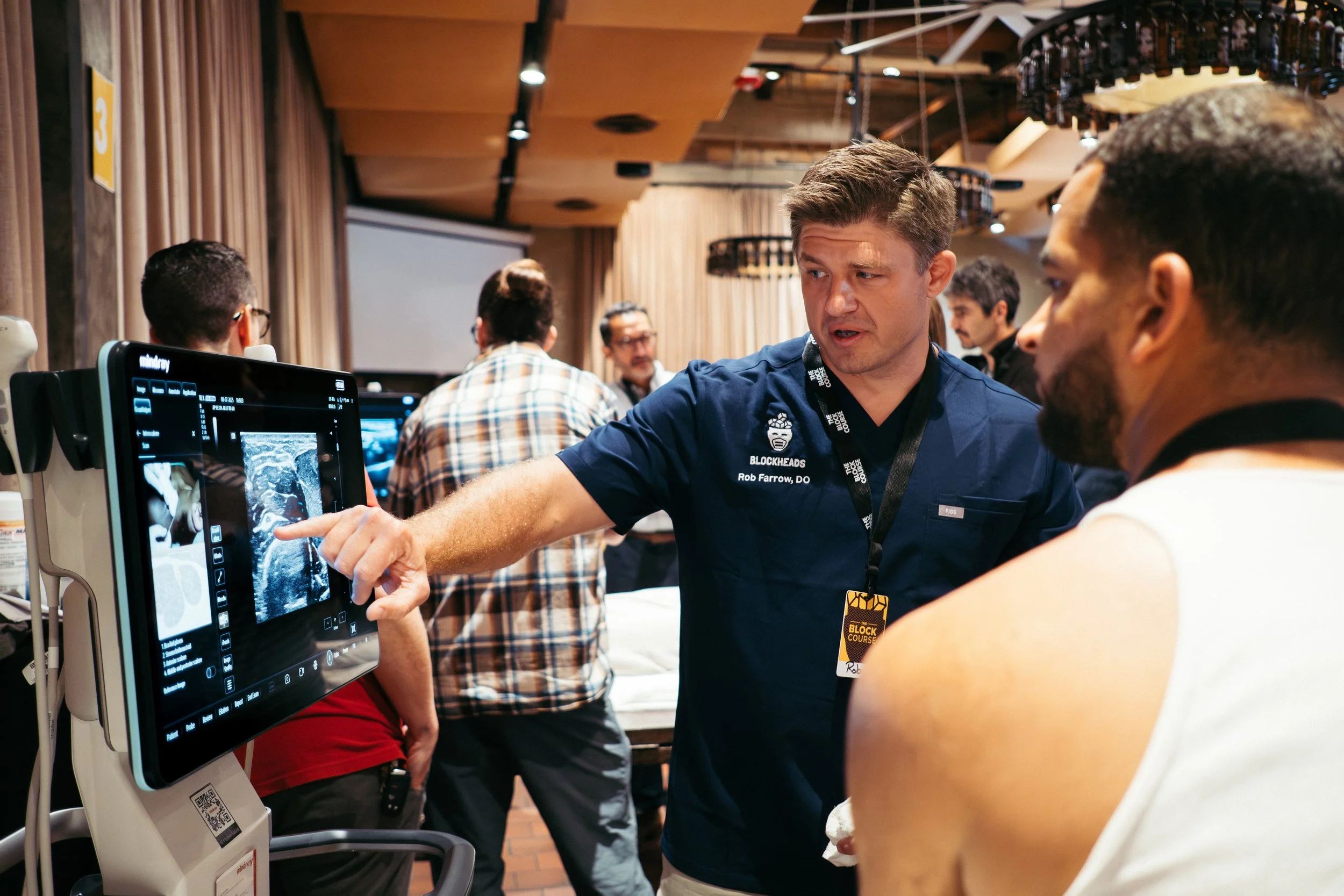
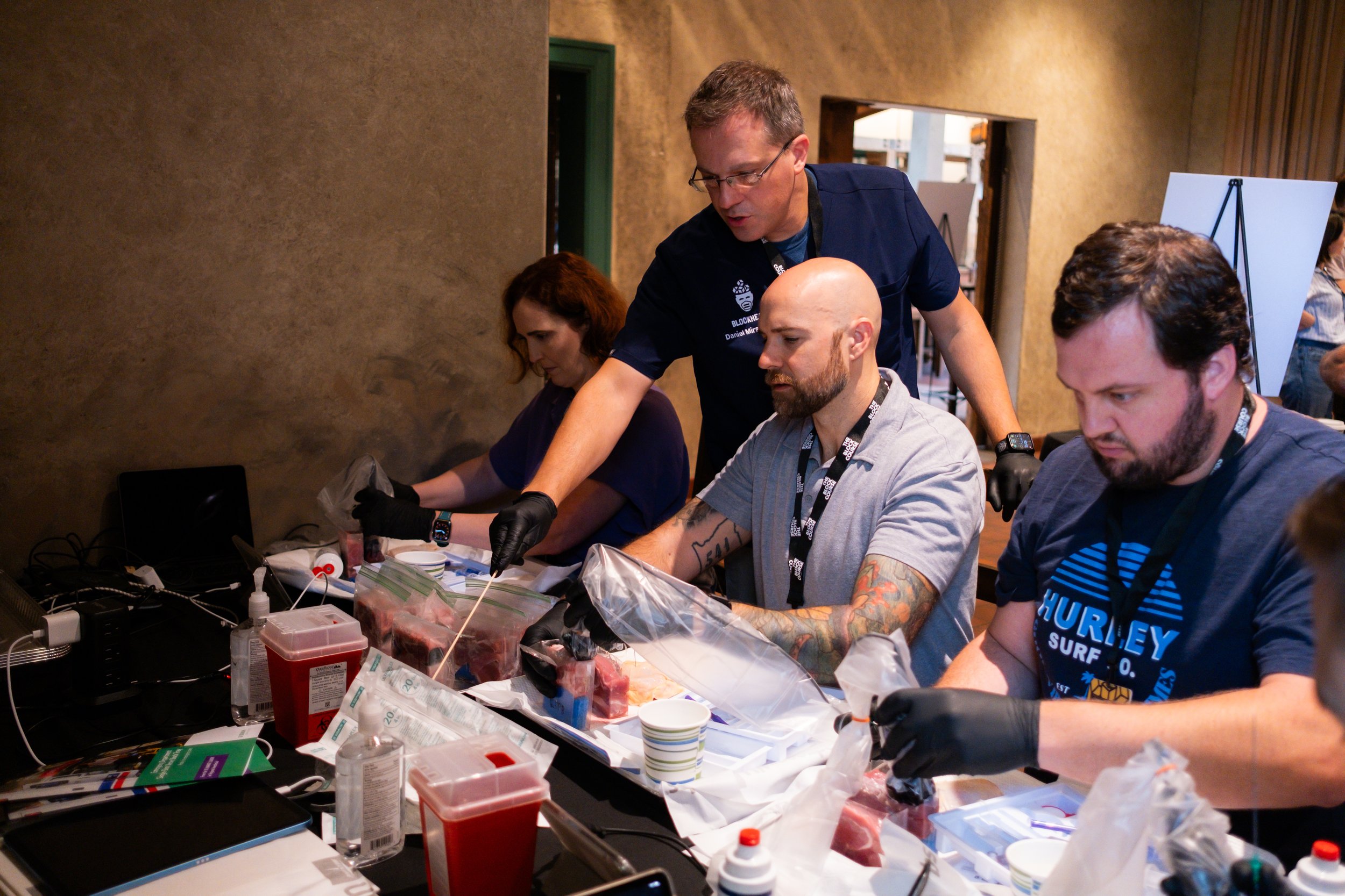
Emergency physicians interested in expanding their use of regional anesthesia can learn these techniques through hands-on training, cadaver labs, and structured educational programs as part of the BlockHeads community.
BlockHeads courses focus on practical ultrasound-guided regional anesthesia techniques that can be safely implemented in the emergency department.
Explore upcoming courses, research opportunities, and educational resources to continue developing your nerve block skills!
If you prefer to learn via a community of like minded physicians be sure to join our next BlockHeads Forum!
A Practical Guide for Emergency Physicians
Ultrasound-guided regional anesthesia is rapidly becoming a core procedural skill in modern emergency medicine. For many common injuries seen in the emergency department — fractures, dislocations, rib injuries, and complex lacerations — nerve blocks can provide rapid, targeted analgesia while reducing reliance on opioids and procedural sedation.
At BlockHeads, we focus on practical techniques that emergency physicians can integrate into everyday clinical practice. The following list highlights 20 ultrasound-guided nerve blocks that are particularly useful in the emergency department, emphasizing approaches that are effective, reproducible, and well suited to the point-of-care ultrasound environment.
Blocks are organized by anatomic region, reflecting common emergency department presentations and the way clinicians typically approach regional anesthesia at the bedside.
Each block has a quick Best for and Clinical Pearl listed. This is not an exhaustive list of ED US-guided nerve blocks but rather a high-yield list of useful blocks to consider as we care for patients.
Be on the lookout for additional educational content coming soon!!
Top 20
Upper Extremity Blocks
Supraclavicular Brachial Plexus Block
Provides dense anesthesia for most procedures distal to the shoulder.
Best for
Forearm fractures
Wrist injuries
Hand procedures
Clinical Pearl
Look for the classic “cluster of grapes” appearance of the brachial plexus lateral to the subclavian artery.
Interscalene Block
Provides excellent analgesia for shoulder pathology.
Best for
Shoulder dislocation reduction
Proximal humerus fractures
Clinical Pearl
Identify the brachial plexus roots between the anterior and middle scalene muscles.
RAPTIR Block (Retroclavicular Approach to the Infraclavicular Region)
A variation of the infraclavicular brachial plexus block that targets the cords of the plexus while improving needle visualization.
Best for
Elbow injuries
Forearm fractures
Wrist and hand procedures
Clinical Pearl
The retroclavicular needle trajectory often provides excellent alignment with the ultrasound beam, improving needle visualization.
Radial Nerve Block — Supracondylar Approach
Provides anesthesia to the dorsal hand and wrist.
Best for
Distal radius fractures
Dorsal hand lacerations
Metacarpal fractures
Clinical Pearl
Start scanning at the mid-humerus where the nerve exits the spiral groove, then track distally toward the supracondylar region.
Median Nerve Block
Provides anesthesia to the palmar hand.
Best for
Palmar hand lacerations
Carpal tunnel procedures
First–third digit injuries
Clinical Pearl
Start scanning distally in the carpal tunnel and track proximally to confirm nerve identity.
Ulnar Nerve Block
Provides anesthesia to the medial hand and fifth digit.
Best for
Fifth digit injuries
Medial hand lacerations
Clinical Pearl
Distally the nerve separates from the ulnar artery into its own fascial plane.
Ultrasound-Guided Flexor Tendon Sheath Block (Ultrasound Guided Digital Block)
Provides targeted analgesia for individual digits through anesthetic deposition within the flexor tendon sheath.
Best for
Finger laceration repair
Finger fracture reduction
Nail bed procedures
Clinical Pearl
Identify the flexor tendon within the tendon sheath on ultrasound and inject small volumes while visualizing spread within the sheath.
Lower Extremity Blocks
Infrainguinal Fascia Iliaca Block
A commonly used technique for hip fracture analgesia.
Best for
Hip fractures
Femoral neck fractures
Clinical Pearl
Ensure the needle tip is deep to the fascia iliaca, with hydrodissection lifting the fascia.
Suprainguinal Fascia Iliaca Block
Allows more reliable spread toward lumbar plexus branches.
Best for
Hip fractures
Acetabular injuries
Clinical Pearl
Position the probe just medial to the ASIS and align it along the iliac crest.
PENG Block (Pericapsular Nerve Group Block)
Targets articular branches supplying the anterior hip capsule.
Best for
Hip fracture analgesia
Hip dislocation reduction
Clinical Pearl
The injection target lies near the iliopubic eminence along the superior pubic ramus.
Proximal Sciatic Nerve Block (Trans-Gluteal Approach)
Provides anesthesia to most of the lower leg and foot.
Best for
Tibia and fibula fractures
Severe lower leg trauma
Extensive distal extremity procedures
Clinical Pearl
The sciatic nerve lies deep to the gluteus maximus between the greater trochanter and ischial tuberosity.
Popliteal Sciatic Nerve Block (Distal Sciatic Nerve Block)
Provides anesthesia to the lower leg, ankle, and foot.
Best for
Ankle fractures
Foot injuries
Distal lower extremity procedures
Clinical Pearl
Perform the block proximal to the bifurcation of the sciatic nerve into the tibial and common peroneal nerves.
Distal Tibial Nerve Block (Posterior Tibial Nerve Block)
Provides anesthesia to the plantar surface of the foot.
Best for
Plantar foot lacerations
Calcaneal injuries
Foreign body removal
Clinical Pearl
The nerve lies posterior to the posterior tibial artery near the medial malleolus.
Saphenous Nerve Block (Adductor Canal Block)
Provides sensory anesthesia to the medial lower leg and ankle.
Best for
Medial ankle injuries
Medial foot lacerations
Clinical Pearl
The nerve is visualized adjacent to the femoral artery beneath the sartorius muscle within the adductor canal.
Truncal Blocks
Serratus Anterior Plane Block
Provides chest wall analgesia.
Best for
Rib fractures
Thoracic trauma
Clinical Pearl
Inject between the latissimus dorsi and serratus anterior muscles.
Erector Spinae Plane Block
Provides analgesia for thoracic pain.
Best for
Rib fractures
Thoracic back pain
Clinical Pearl
Identify the transverse process beneath the erector spinae muscle.
Transversus Abdominis Plane (TAP) Block
Provides analgesia to the anterior abdominal wall.
Best for
Abdominal wall pain
Laceration repair
Clinical Pearl
Inject between the internal oblique and transversus abdominis muscles.
Rectus Sheath Block
Provides midline abdominal wall analgesia.
Best for
Umbilical hernia reduction
Periumbilical procedures
Clinical Pearl
Local anesthetic spreads within the posterior rectus sheath.
Head, Neck, and Shoulder Blocks
Superficial Cervical Plexus Block
Provides sensory blockade to the lateral neck and clavicular region.
Best for
Clavicle fractures
Neck lacerations
Cervical soft tissue procedures
Clinical Pearl
The cervical plexus branches emerge along the posterior border of the sternocleidomastoid muscle.
Clavipectoral Fascial Plane Block
Targets the sensory branches supplying the clavicle through local anesthetic deposition within the clavipectoral fascia.
Best for
Clavicle fractures
Clavicle procedures
Clinical Pearl
Inject local anesthetic between the clavipectoral fascia and clavicle near the fracture site.