* UGRA Simulation*
DIY - “Pocket” UGRA Meat Model Design
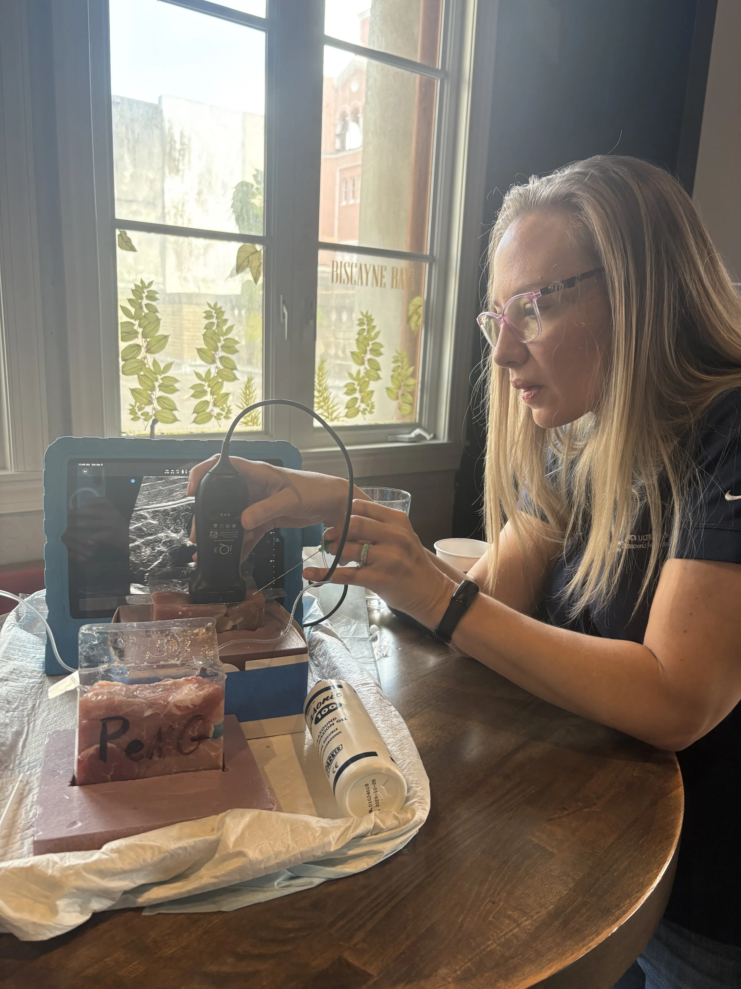
Created by: Dr. Dan Mirch, DO
Meat models for nerve block training are an effective and economical alternative to commercially available simulators. While many commercial models do not allow for the injection of fluid or the demonstration of fascial plane separation, our model addresses these limitations.
Although our models require hands-on manufacturing and a foundational understanding of anatomy, their cost-savings is a significant advantage, which we pass on to our learners. We assert that these models are superior to current alternatives because they:
* Clearly mimic anatomical structures.
* Feature a realistic tissue appearance.
* Withstand multiple needle punctures without degradation.
* Enable realistic fascial plane separation, allowing for the correction of improper injection techniques.
** Hydrodissection can be performed using either a spinal needle or a nerve block needle injecting tap water. **

-
Clear Plastic Box: The clear plastic box serves as a transparent container for visualization of the internal model structures and holds all the components together to prevent model degradation during use. (BENECREAT 30PCS Clear Gift Boxes 4x4x1.2 inch Rectangle PVC Clear Wedding Favour Boxes for Candy Chocolate)
Sponge: The sponge elevates the placement of meat cuts in the box and appears to have a hyperechoic rim which may also mimic bone underneath if needed
Muscle Groups: Boneless pork tenderloins are cut into various shapes and sizes to mimic different muscle groups. The cuts are made to simulate the size, shape, and direction of muscle fibers.
Fascial Planes: Fascial planes are created by layering the muscle cuts on top of each other. No binding ingredient is needed. The presence of natural fascia on the meat cuts enhance the realism of the fascial planes.
Nerves: Pieces of bulky yarn soaked in ultrasound gel simulate nerves. The gel soaked yarn can be folded over multiple times to create a larger appearing nerve. When layered linearly, the yarn also demonstrates anisotropic properties yielding a realistic sonographic appearance. Gel filled regular sized straws are also used to simulate the anechoic interscalene brachial plexus.
Vessels: Straws are cut to 1 inch length and filled with ultrasound gel to simulate large vessels.
Bone: Take out containers (Fold Pak 15 count 16 oz Pagoda Wire Handle Chinese Takeout Box) are cut into strips to mimic bone in the PeNG model. Either whisky rocks (1”x1” or 0.79”x0.79” rocks) or pork spare ribs can be used to mimic bone. If using spare rib bones, the ribs are cut with a meat cleaver into 1” width and surrounding meat removed with trauma shears
-
Forearm Model
Components:
Sponge: 4”x2” piece.
Transverse cut of pork tenderloin: mimics flexor digitorum profundus (FDP).
Gel soaked yarn: mimics median nerve.
Transverse cut of pork tenderloin: mimics flexor digitorum superficialis (FDS).
Assembly: Place the sponge in the bottom of the box. Insert the FDP and sandwich the median nerve between the FDP and the FDS on top. A shallow cut can be made in one of the pork tenderloins to house the median nerve.
Interscalene Brachial Plexus (ISBP) Model
Components:
Sponge: 4”x2” piece.
Transverse cut of pork tenderloin cut in half but not all the way through: mimics anterior scalene (AS) and middle scalene (MS) muscles.
Three gel filled regular sized straws: mimics C5, C6, and C7 nerve roots of the interscalene brachial plexus.
Gel filled boba straw: mimics carotid artery.
Thin triangular slice of meat: mimics sternocleidomastoid muscle (SCM).
Small filler meat pieces: used to fill up the area underneath the carotid artery.
Assembly:
Place the sponge in the bottom of the box. Sandwich the three gel filled straws vertically between the AS and MS muscles. Place this structure onto the sponge and fill in remaining adjacent spaces with filler meat. Place the carotid artery adjacent to the scalene muscles. Place the SCM on top of the model.
-
Erector Spinae Plane Block (ESPB) Model
Components:
Sponge: 4”x2” piece with two cutouts around 0.5” deep to fit the whisky rocks.
Two whisky rocks or 1” cuts of rib bone: mimics ribs.
Three 0.5”x1” thick filler meat pieces: fill in space between whisky rocks.
4”x0.25” Longitudinal cut of pork tenderloin: mimics erector spinae muscle.
4”x1” thick cut of pork tenderloin: mimics other muscles and subcutaneous tissue above the erector spinae.
Assembly:
Place the sponge in the bottom of the box and place the two whisky rocks into the cutouts into the sponge to prevent the rocks from moving. Alternatively, pork spare rib bones can be cut in 1” pieces to mimic ribs. Fill the empty space between the whisky rocks with filler meat. Layer the thin 0.25” cut of pork tenderloin on top, followed by the 1” cut of pork tenderloin.
Serratus Anterior Plane Block (SAPB) Model
Components:
Sponge: 4”x2” piece with cutouts same as ESP above.
Two whisky rocks or 1” cuts of rib bone: mimics ribs.
Three 0.5”x1” thick filler meat pieces: fill in space between whisky rocks.
4”x0.5” thick longitudinal cuts of pork tenderloin: mimics serratus anterior muscle.
3”x0.5” thick triangular cut of pork tenderloin: mimics lateral edge of latissimus dorsi muscle.
4” layer of filler meat on top: mimics subcutaneous tissue
Assembly:
Place the sponge, whisky rocks, and filler meat similarly to the ESPB model. Layer the 0.5” thick meat on top. Layer the triangular piece to represent the tapered edge of the latissimus dorsi muscle. Lastly, place a layer of filler meat on top for the subcutaneous tissue.
-
Fascia Iliaca Compartment Block (FICB) Model
Components:
Sponge: 4”x2” piece.
Transverse cut of pork tenderloin with small cut in the meat: mimics iliopsoas muscle with a small cut to house the femoral nerve.
Large triangular cut of meat: mimics sartorius muscle.
Gel soaked yarn: mimics femoral nerve.
Gel filled boba straw: mimics femoral artery.
Assembly:
After placing the sponge in the bottom of the box, place the iliopsoas on top of the sponge and wedge the femoral nerve into the small cut placed into the iliopsoas. Then place the sartorius muscle on top to one side and the femoral artery to the other side. Filler meat can be used to support the femoral artery and to fill the subcutaneous space.
Pericapsular Nerve Group (PeNG) Model
Components:
Sponge: 4”x2” cut with a “L” shape
Takeout container cut into a 1”x5” strip taped to box: mimics the anterior inferior iliac spine (AIIS) and iliopubic eminence (IPE).
Transverse cut of pork tenderloin with small cut in the meat: mimics psoas muscle with a small cut to house the psoas tendon.
Transverse cut of pork tenderloin: mimics iliacus muscle.
Gel soaked yarn: mimics psoas tendon.
Gel filled boba straw: mimics femoral artery.
Filler meat: mimics subcutaneous tissue.
Assembly:
Insert the sponge and then layer the strip of takeout container on top, bending the takeout container to create the “L” shape. Next, place the gel soaked yarn into the small cut placed in the psoas muscle to represent the psoas tendon. Place the psoas with the tendon facing downwards. Place the iliacus muscle to the side of the psoas muscle closest to the AIIS with the femoral artery to the other side. Cover the model with filler meat to represent subcutaneous tissue.
Posterior Tibial Model
Components:
Sponge: cut into a right triangle with 4”x2.5” sides 90 degrees to each other to mimic the medial malleolus.
Takeout container: 4.5”x1” strip to mimic bony cortex of medial malleolus (optional).
Three gel filled regular sized straws: mimics the posterior tibial artery and veins.
Gel soaked yarn: mimics posterior tibial nerve.
Two transverse cuts of pork tenderloin: one cut mimics the flexor hallucis longus (FHL) muscle and the other cut mimics the tibialis posterior (TP) and flexor digitorum longus (FDL) muscles together.
One 0.5” slice of meat of uniform thickness: mimics subcutaneous tissue.
Assembly:
Wedge the triangular sponge to the bottom of the box. Lay the takeout container strip on top (optional as the sponge itself will already have a hyperechoic rim which mimics bone). Place the two transverse cuts of meat on top of the sponge. In between the two meat pieces, wedge two regular sized gel filled straws next to each other. Wedge the gel soaked yarn right next to the straws on the side closest to the lowest slope. Cover model with meat of uniform thickness.
Transgluteal Sciatic Nerve Model
Components:
0.5” slice of meat of uniform thickness: mimics subcutaneous tissue.
2”x1” piece of filler meat: mimics quadratus femoris and gluteus maximus.
Gel soaked yarn: mimics sciatic nerve.
4”x1.5” piece of meat: mimics gluteus maximus.
Two pieces of sponge cut out to 1” width and 2.5” height: mimics greater trochanter of femur and ischial tuberosity.
Assembly: Wedge the two pieces of sponge to either side of the box. Insert the 2”x1” meat in between the sponges. Make a small cut on the second pork tenderloin piece to house the sciatic nerve and place this piece nerve side down.